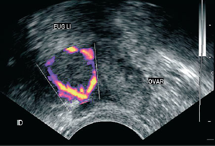

The gel allows sound waves to travel back and forth between the transducer and the area under examination. The technologist applies a small amount of gel to the area under examination and places the transducer there. The same principles apply to sonar used by boats and submarines. The transducer sends out inaudible, high-frequency sound waves into the body and listens for the returning echoes. Some exams may use different transducers (with different capabilities) during a single exam. The transducer is a small hand-held device that resembles a microphone. Ultrasound machines consist of a computer console, video monitor and an attached transducer. In children, pelvic ultrasound can help evaluate: In men and women, a pelvic ultrasound exam can help identify: See the Prostate Ultrasound page for more information. Transrectal ultrasound, a special study usually done to provide detailed evaluation of the prostate gland, involves inserting a specialized ultrasound transducer into a man's rectum. In men, a pelvic ultrasound is used to evaluate the: See the Sonohysterography page for more information. In this setting, three-dimensional ultrasound provides information about the outer contour of the uterus and about uterine irregularities, as well as information about the fallopian tubes and ovaries. Some physicians also use 3-D ultrasound or sonohysterography or Hysterosalpingo-Contrast-Sonography (H圜oSy) for patients with infertility. cancer, especially in patients with abnormal uterine bleeding.


These exams are typically performed to detect: Three-dimensional (3-D) ultrasound permits evaluation of the uterus and ovaries in planes that cannot be imaged directly. Sonohysterography allows for a more in-depth investigation of the uterine cavity and Hysterosalpingo-Contrast-Sonography (H圜oSy) allows for assessment of patency of the fallopian tubes. Transvaginal ultrasound also evaluates the myometrium (muscular walls of the uterus) and the fallopian tubes. palpable masses such as ovarian cysts and uterine fibroidsĪ transvaginal ultrasound is usually performed to view the endometrium (the lining of the uterus) and the ovaries.Ultrasound examinations can help diagnose symptoms experienced by women such as: See the Obstetrical Ultrasound page for more information. Pelvic ultrasound exams are also used to monitor the health and development of an embryo or fetus during pregnancy.

In women, a pelvic ultrasound is most often performed to evaluate the: What are some common uses of the procedure? It allows the doctor to see and evaluate blood flow through arteries and veins in the body.


 0 kommentar(er)
